
Introduction
If you’ve noticed a soft, tender swelling sitting directly on top of your kneecap, and kneeling has become painful, you may have prepatellar bursitis.
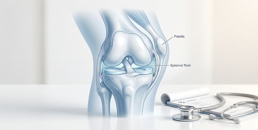
The prepatellar bursa is a small, flattened sac that lies just beneath the skin at the front of your kneecap. Its job is to let the skin glide smoothly over the bone underneath. Normally it holds barely a trace of fluid and you’d never know it was there. When it becomes irritated, it fills up — and because it sits so close to the surface, the resulting swelling is often strikingly visible, frequently described as an egg or a small ball resting on the front of the knee.
One point is worth clearing up straight away, because it causes a lot of confusion. Prepatellar bursitis is not the same as fluid on the knee joint. The bursa sits outside the joint capsule entirely, so this is a problem in front of the knee rather than inside it. The two conditions look different, feel different, and are treated differently.
Prepatellar bursitis turns up most often in people who kneel for a living — flooring fitters, plumbers, roofers, gardeners, and cleaners — which is why it has long been known as housemaid’s knee. It also follows a hard knock to the knee, and can be triggered by gout or an underlying inflammatory condition.
It also has one feature that sets it apart from most knee problems. Because only a thin layer of skin separates the bursa from the outside world, a minor cut or graze over the kneecap can let bacteria in. A meaningful share of cases turn out to be infected, and infected bursitis needs antibiotics rather than rest and ice. Telling the two apart is the single most important question in this condition, and it’s one you can’t reliably answer by looking.
This article covers the anatomy, what causes prepatellar bursitis, how to recognize it, how doctors distinguish an infected bursa from an inflamed one, and the treatment options for each.
What Is Prepatellar Bursitis?
Prepatellar (kneecap) bursitis is a common condition that may occur in everyone but is more common among plumbers, coal miners, and footballers. The knees have a bursa sac, a fluid-filled sac that reduces the friction between the tissues.
If you’re in a job where you spend more time putting more pressure on your knees, you’re more likely to get this. Though the pain caused by prepatellar bursitis reduces on its own, if it’s severe, you may have to consult a doctor and get it treated immediately. So, if you notice any pain in the knee, it may be due to this.
What Is A Bursa?
A bursa is a small, flattened sac lined with synovial membrane and containing a thin film of fluid. Bursae sit at points where tissues rub against one another — typically where skin, tendon, or muscle passes over bone — and allow those surfaces to glide rather than catch. The body contains roughly 150 of them.
A healthy bursa is almost flat, holding only a trace of fluid. It becomes noticeable only when it is irritated and fills.
Where the prepatellar bursa sits
The prepatellar bursa lies directly in front of the kneecap, between the skin and the front surface of the patella. It is superficial, meaning it sits just beneath the skin with very little tissue covering it.
Two features of that position explain almost everything about the condition:
It is exposed. With only skin over it, the bursa absorbs direct impact and sustained pressure whenever the knee is knelt on or struck. Nothing cushions it.
It is close to the surface. Because only a thin layer of skin separates it from the outside world, a cut, graze, scratch, or insect bite over the kneecap can allow bacteria to reach the bursa directly. This is why infection is far more common in prepatellar bursitis than in most other joint-region problems.
Why this is not the same as a knee joint problem
The knee joint itself is enclosed in a capsule containing the cartilage surfaces, ligaments, and menisci. The prepatellar bursa sits outside that capsule and, in most people, does not communicate with it.
Practical consequences:
- Prepatellar bursitis produces swelling that is in front of the kneecap and looks localized and superficial, often described as an egg or golf ball sitting on the knee
- Knee joint effusion produces swelling that is diffuse across the whole joint, filling the hollows around the kneecap
- Prepatellar bursitis usually leaves knee bending largely intact, though kneeling is painful; a joint effusion typically restricts bending and straightening considerably
- Infection of the bursa is a serious but generally localized problem; infection inside the joint, septic arthritis, is a surgical emergency that destroys cartilage
The other bursae around the knee
Several bursae sit around the knee and are sometimes confused with one another:
- Suprapatellar bursa — above the kneecap, and unlike the prepatellar bursa it does connect with the knee joint in most adults
- Superficial infrapatellar bursa — below the kneecap, in front of the patellar tendon
- Deep infrapatellar bursa — behind the patellar tendon
- Pes anserine bursa — on the inner side of the knee, a few inches below the joint line, and a common cause of inner knee pain

Types Of Prepatellar Bursitis
Prepatellar bursitis is classified in two ways at once, and both matter.
By time course
Acute prepatellar bursitis develops over hours to days, typically after a direct blow or a short period of intense kneeling. The bursa fills rapidly, and the swelling is often tender and sometimes warm.
Chronic prepatellar bursitis develops gradually over weeks to months, usually from repeated kneeling. The bursa wall thickens over time, and the swelling may be firmer, less tender, and more persistent. Chronic cases can flare acutely.
By cause
This is the more important distinction.
Aseptic (non-infected) bursitis accounts for most cases. The bursa is inflamed from trauma, repetitive pressure, crystal deposits, or an underlying inflammatory condition, but no bacteria are present.
Septic (infected) bursitis occurs when bacteria enter the bursa, most often through a break in the skin over the kneecap. It is considerably more common in the prepatellar bursa than in deeper sites, precisely because the bursa lies so close to the surface. In published case series, a substantial minority of prepatellar bursitis cases — commonly cited as up to around a third — turn out to be septic.
That proportion is the single most important fact about this condition, and it is why the septic-versus-aseptic question runs through the rest of this article. The two are treated completely differently, and they cannot reliably be told apart by how the knee looks.
Causes of Prepatellar Bursitis
Repetitive kneeling
The most common cause. Sustained or repeated pressure on the front of the kneecap irritates the bursa, which responds by producing fluid and, over time, thickening its wall.
Occupations and activities with elevated risk include flooring and carpet fitting, plumbing, roofing, tiling, mining, construction, gardening, cleaning, and any work involving prolonged floor-level tasks. Wrestling, volleyball, and basketball carry higher rates among sports.
The risk relates to total kneeling time and whether any padding is used, rather than to occupation as such.
Direct trauma
A single hard blow to the front of the knee — a fall onto a hard surface, a collision, an impact in sport — can cause bleeding or rapid fluid accumulation within the bursa. Swelling after a direct impact often appears within hours.
Infection
Bacteria reach the bursa through a break in the overlying skin. That break can be minor and easy to overlook: a graze, a small cut, a scratch, an insect bite, an area of chapped skin, or a shaving nick.
Staphylococcus aureus is the organism most often responsible, reflecting its presence on normal skin.
Risk is higher in people with diabetes, a weakened immune system, alcohol dependence, existing skin conditions such as eczema over the knee, kidney disease, or a previous episode of bursitis in the same site.
Septic bursitis can also develop without any remembered skin injury, which is one reason it is missed.
Gout and other crystal conditions
Uric acid crystals can deposit in a bursa just as they do in a joint. Gout affecting the prepatellar bursa produces sudden, intensely painful swelling with marked redness and heat — a presentation that closely mimics infection and cannot be separated from it without testing the fluid.
Calcium pyrophosphate crystals (pseudogout) can do the same, more often in older adults.
People with established gout, a history of gout attacks elsewhere, or raised uric acid are at higher risk.
Inflammatory arthritis
Rheumatoid arthritis and other inflammatory conditions can involve bursae as well as joints, producing bursitis as part of a wider disease process. This tends to be less abrupt than infection or gout and is usually accompanied by symptoms elsewhere.
A note on osteoarthritis
Osteoarthritis of the knee does not directly cause prepatellar bursitis — they affect different structures. There is an indirect link, in that knee osteoarthritis can change how a person kneels and moves, potentially increasing pressure on the bursa, and both conditions are more common with higher body weight. The existing article’s suggestion that arthritis pain “worsens and causes bursitis” overstates a relationship that is indirect at best.
Prepatellar Bursitis Symptoms

The characteristic picture
- A localized swelling directly over the kneecap, often strikingly well-defined and frequently compared to an egg or a small ball sitting on the front of the knee
- Pain on kneeling, which is usually the symptom that brings people in, and which can be severe even when the knee is otherwise comfortable
- Tenderness when the swelling is pressed
- A squashy or fluid-filled feel to the swelling in acute cases, or a firmer, more rubbery feel in chronic ones
- Skin changes over the swelling in long-standing cases, including thickening or callus
What is usually preserved
This is as informative as the symptoms themselves. In uncomplicated prepatellar bursitis:
- Knee bending is largely preserved. It may be uncomfortable at the extreme of flexion because the skin stretches over the swelling, but the range is not usually greatly restricted
- Walking is generally possible, though kneeling is not
- The knee joint itself is not usually tender along the joint line
Marked restriction of knee movement, pain deep within the joint, or inability to bear weight suggests the problem is not confined to the bursa, and points toward the joint itself.
Symptoms that suggest infection
These change the urgency completely:
- Fever or chills
- Redness spreading beyond the swelling onto the surrounding skin
- Marked warmth compared with the other knee
- Severe tenderness, often out of proportion to the size of the swelling
- Rapidly worsening pain and swelling over hours
- A visible break in the skin over the kneecap — a cut, graze, or bite
- Feeling generally unwell
Anyone with a swollen kneecap and any of these features needs same-day medical assessment. Gout can produce a very similar picture, and that is a reason for prompt assessment rather than reassurance, since the two cannot be distinguished by appearance.
Septic vs Aseptic: How to Tell the Difference
This is the central clinical question in prepatellar bursitis, and it deserves its own section because the article’s readers will mostly be trying to answer it about their own knee.
Features that point each way
| Feature | More typical of aseptic | More typical of septic |
|---|---|---|
| Onset | Gradual, or hours after a knock | Often rapid, over 24 to 48 hours |
| Fever | Absent | Present in many cases |
| Skin redness | Mild or absent | Marked, often spreading beyond the swelling |
| Warmth | Mild | Pronounced |
| Tenderness | Moderate | Severe, often disproportionate |
| Skin break over the knee | Usually none | Frequently present, sometimes minor |
| Feeling unwell | No | Often |
| Preceding cause | Kneeling or trauma | May follow a minor skin injury |
The honest limitation
These features overlap, and none of them is reliable on its own. Septic bursitis can present without fever, particularly early or in people on anti-inflammatory medication. Gout can produce redness, heat, and fever that look exactly like infection. Aseptic bursitis following a hard blow can be red, warm, and very tender.
Studies looking at how accurately clinicians distinguish septic from aseptic bursitis on examination alone consistently find the two are frequently confused. If experienced clinicians cannot reliably separate them by looking, a reader cannot do so at home.
The practical conclusion: a swollen, red, warm, or painful kneecap should be assessed rather than self-managed. The definitive answer comes from testing fluid drawn from the bursa, not from observation.
How Prepatellar Bursitis Is Diagnosed
History and examination
A doctor will ask about kneeling at work or in sport, recent injury or blows to the knee, any cuts or grazes over the kneecap, how quickly the swelling appeared, whether there has been fever, previous episodes, gout history, and conditions such as diabetes.
The examination assesses the location and character of the swelling, skin temperature compared with the other knee, the extent of any redness, whether the skin is intact, knee range of motion, and whether the knee joint itself is tender. Nearby lymph nodes may be checked.
A key part of the examination is confirming that the swelling is in front of the kneecap rather than within the joint.
Bursal aspiration
Drawing fluid from the bursa with a needle is the definitive test, and it is the only reliable way to separate septic from aseptic bursitis.
The fluid is examined for:
- Appearance — clear or straw-colored in mild aseptic cases, blood-stained after trauma, cloudy or frankly purulent in infection
- White cell count — low in aseptic bursitis, markedly raised in infection
- Gram stain and culture — identifies bacteria and determines which antibiotics will be effective
- Crystals under polarized microscopy — confirms gout or pseudogout
Aspiration also relieves pressure and pain when the swelling is large.
Because the prepatellar bursa is superficial, the procedure is quick and done under local anesthetic. It is generally avoided through visibly infected or broken skin, to avoid carrying bacteria inward.
Blood tests
Inflammatory markers (ESR, CRP), a full blood count, and sometimes blood cultures may be taken where infection is suspected. Uric acid may be checked if gout is a consideration, though levels can be normal during an attack.
Imaging
Imaging is not usually needed for a clear-cut case.
- X-ray is used if a fracture is suspected after trauma, or to look for a foreign body
- Ultrasound confirms fluid within the bursa, distinguishes bursal from joint swelling, assesses bursal wall thickening, and can guide aspiration
- MRI is reserved for unclear cases, or where deeper infection or another diagnosis is suspected
Conditions that can look similar
- Knee joint effusion
- Cellulitis of the skin over the knee, without bursal involvement
- Infrapatellar bursitis, sitting below rather than over the kneecap
- Gout in the knee joint itself
- Patellar fracture following trauma
- A cyst or, rarely, a soft tissue tumor

Prepatellar Bursitis Treatment
Treatment depends entirely on whether the bursitis is septic or aseptic, which is why that question is settled first.
Treatment of septic bursitis
Septic bursitis requires medical treatment. It does not resolve with rest and ice.
- Aspiration of the infected fluid, both to obtain a sample and to reduce the bacterial load
- Antibiotics, chosen to cover the likely organisms and then adjusted once culture results identify what is present. Most people are treated with oral antibiotics as outpatients; more severe cases, or people with weakened immunity, may need intravenous treatment in hospital
- Repeat aspiration if fluid reaccumulates
- Surgical drainage or removal of the bursa if the infection does not settle with antibiotics and aspiration
Antibiotic choice and duration are individual clinical decisions made after assessment, and depend on the organism, its resistance pattern, and the person’s medical history.
Corticosteroid injection must not be given into an infected bursa. This is one of the main practical reasons infection is excluded before any injection is considered.
Treatment of aseptic bursitis
Most cases settle with conservative measures over several weeks.
- Activity modification is the foundation. Avoiding kneeling is the single most effective step, and it is more important than rest in general — walking and normal daily activity are usually fine and preferable to inactivity, which causes stiffness and muscle loss.
- Ice applied for 15 to 20 minutes at a time, with a cloth between the pack and the skin, several times daily during the acute phase.
- Compression using an elastic sleeve or bandage, firm but not tight enough to affect circulation.
- Elevation where practical.
- NSAIDs such as ibuprofen or naproxen, for pain and inflammation. They are not suitable for everyone, particularly people with stomach, kidney, or cardiovascular problems. Topical NSAID gel is an option given how superficial the bursa is.
- Aspiration may be performed if the swelling is large and uncomfortable, and it has the advantage of providing fluid for testing at the same time.
- Corticosteroid injection is sometimes used for persistent aseptic bursitis, but it is more contentious here than at other sites. The bursa’s superficial position means injection carries risks of skin thinning, pigment change, and introducing infection. Many clinicians use it sparingly and only after infection has been definitively excluded.
- Knee padding during any unavoidable kneeling.
Surgery
Surgery is uncommon and reserved for cases that do not respond to everything else.
Bursectomy, removal of the bursa, may be considered for chronic bursitis that persists despite months of conservative management, for recurrent episodes, or for septic bursitis that fails to clear.
It can be performed open or endoscopically. Endoscopic approaches have been described with the aim of faster recovery and fewer wound problems, since the skin over the kneecap heals slowly and wound complications are a recognized issue with open surgery at this site.
Recovery involves protecting the wound, avoiding kneeling during healing, and a graded return to activity.
Conservative Management and Recovery
Realistic timelines
Acute aseptic bursitis from a single knock often settles within two to six weeks with activity modification. Chronic bursitis from years of kneeling takes longer and may not resolve completely, particularly where the bursal wall has thickened. Septic bursitis usually improves within days of starting appropriate antibiotics, though full resolution takes longer. Recurrence is common where the original cause — usually kneeling — continues unchanged.
Getting back to work
This is the practical problem for most people with this condition, since it is largely an occupational one. Useful steps include:
- Good quality knee pads, gel or foam, worn consistently rather than occasionally
- A kneeling mat or board for floor-level work
- Changing position regularly rather than kneeling continuously
- Alternating knees where the task allows
- Working from a low stool instead of kneeling where possible
- Keeping the skin over the knee intact, since breaks in the skin are the route to infection
Strength and movement
Prolonged avoidance of the knee causes quadriceps weakness, so gentle strengthening once the acute phase has passed helps restore normal function. A physical therapist can advise, particularly for chronic or recurrent cases.
Joint and connective tissue support
Some people use supplements as part of a longer-term routine for joint comfort and mobility. It is worth being precise about the limits here, because prepatellar bursitis is a condition of the bursa — a fluid-filled sac under the skin — rather than of the knee joint, its cartilage, or its connective tissue. Supplements do not resolve an inflamed bursa, do not treat infection, crystal conditions, or trauma, and are not a substitute for medical assessment. Given how often prepatellar bursitis turns out to be septic, a swollen kneecap always needs its cause established first.
Where someone is managing knee osteoarthritis alongside occupational knee strain, and the bursitis has been assessed and its cause identified, combination formulas such as Flexoplex Joint Support bring together ingredients commonly used to support joint comfort, flexibility, and everyday mobility.
Supplements can interact with prescription medication, including anticoagulants and some arthritis treatments. Anyone considering them should speak with their doctor or pharmacist first.
Prevention
Prevention centers on reducing pressure on the front of the kneecap and protecting the skin over it.
- Wear knee pads for any kneeling work, and choose ones that are comfortable enough to wear consistently
- Use a kneeling mat for gardening and floor-level tasks
- Take regular breaks from kneeling, standing and moving every 15 to 20 minutes where practical
- Kneel on softer surfaces where there is a choice
- Protect the skin over the knee, and clean and cover any cuts or grazes promptly
- Wear protective equipment in sports involving falls or collisions on hard surfaces
- Maintain a healthy weight, which reduces load through the knee generally
- Address recurrent episodes rather than repeatedly managing them at home, since recurrence usually means the underlying pressure has not changed
Frequently Asked Questions
Q: Can I walk with prepatellar bursitis?
Yes, in most cases. Walking is usually fine and is better than prolonged rest, which causes stiffness and muscle weakness. What needs avoiding is kneeling, and any activity that puts direct pressure on the front of the kneecap. If walking itself is painful, or you cannot bear weight, that suggests the problem may not be confined to the bursa and should be assessed.
Q: How do I know if my bursitis is infected?
Fever, spreading redness, marked warmth, severe tenderness, rapidly worsening swelling, or a cut or graze over the kneecap all point toward infection. But septic and aseptic bursitis overlap considerably, and even experienced clinicians frequently cannot separate them on examination alone. The only reliable answer comes from testing fluid drawn from the bursa, so a red, hot, or severely painful swollen kneecap should be assessed the same day.
Q: Is prepatellar bursitis the same as water on the knee?
No, though the two are often confused. “Water on the knee” usually refers to fluid inside the knee joint, which is joint effusion. Prepatellar bursitis is fluid in a sac in front of the kneecap, outside the joint. Bursitis produces a localized lump over the kneecap; joint effusion produces diffuse swelling across the whole joint and restricts movement more.
Q: How long does prepatellar bursitis take to heal?
Acute cases from a single injury often settle within two to six weeks with activity modification. Chronic cases from prolonged kneeling take longer and may not fully resolve while the kneeling continues. Septic bursitis usually starts improving within days of appropriate antibiotics.
Q: Can prepatellar bursitis come back?
Yes, and recurrence is common when the original cause continues. If the swelling keeps returning, the practical question is what is still putting pressure on the knee, rather than how to treat each episode.
Q: Should I drain it myself?
No. Draining a bursa at home risks introducing bacteria into a space that is already vulnerable, and it discards the fluid that would have identified the cause. Aspiration should be done under sterile conditions by a healthcare professional.
Q: Do I need antibiotics for prepatellar bursitis?
Only if it is infected. Antibiotics do nothing for aseptic bursitis, and taking them unnecessarily carries side effects and contributes to antibiotic resistance. Severity is not the deciding factor — a large, very painful aseptic bursitis still does not need antibiotics, while a milder septic one does.
Q: Can gout cause a swollen kneecap?
Yes. Uric acid crystals can deposit in the bursa as well as in joints, producing sudden, intensely painful swelling with redness and heat. It closely resembles infection and is distinguished by examining the fluid under a microscope.
Conclusion
On the whole, prepatellar bursitis is a common condition that is treatable and occurs in people whose work involves putting pressure on the knees. Sometimes, accidents or hard blows to the knee can cause this, which can be severe if it is infected.
References
- https://www.ncbi.nlm.nih.gov/books/NBK557508/
- https://pmc.ncbi.nlm.nih.gov/articles/PMC3496084/
- https://www.ncbi.nlm.nih.gov/books/NBK441868/
- https://www.niddk.nih.gov/about-niddk/research-areas/kidney-disease
- https://pmc.ncbi.nlm.nih.gov/articles/PMC5512152/