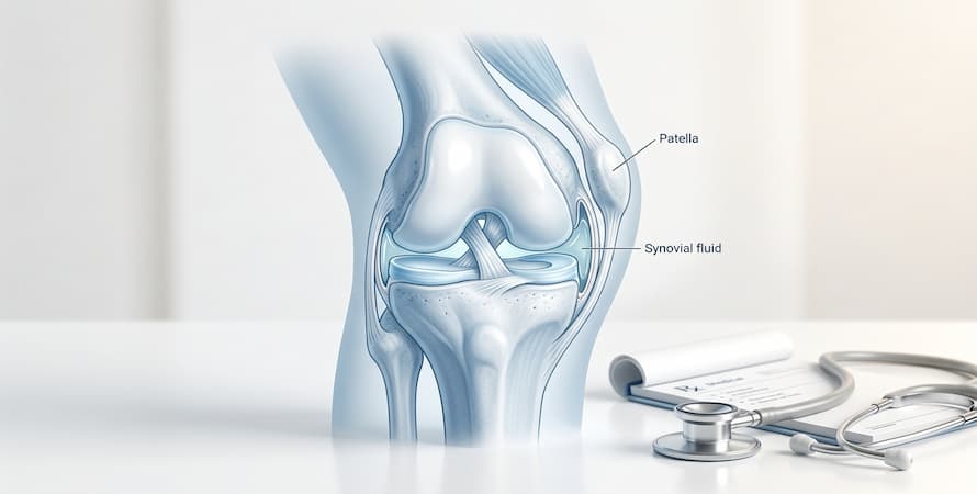

The Definition Of Joint Dislocation And How Common The Injury Is
A joint dislocation is defined as the separation of two bones at the point where they meet in a joint. Your joints are the systems, which are in place to connect bones, so when this connection is interrupted, that is when a dislocation has occurred.
Joint dislocation is common as it may occur in almost any joint in the body. They are also caused by several factors, which is another reason they can happen so frequently.
In fact, one of the most common types of dislocations occurs at the shoulder. It is one of the most common types of dislocation because the shoulder allows the arm to move in many directions, unlike the knee or ankle.
What Causes Joint Dislocation?
There are several factors that contribute to joint dislocation. Of course, regular physical activity and accidents while playing sports, working out, or even being on the job can cause a dislocation with one false move.
Any sort of trauma or impact that pops the bone out of place may cause joint dislocation. For this reason, car accidents are another common cause of this type of injury. However, other factors play into the likelihood that you may suffer, such as dislocation.
Weakened tendons and muscles can also increase your risk of joint dislocation. The muscles and tendons around the joint support it and make sure the bones are protected. When the muscles and tendons become weak due to age, malnutrition, or lack of exercise, it is more likely that dislocation will occur at some point.
Types of Dislocation by Joint
A dislocation happens when the bones forming a joint are forced completely out of their normal position. When they are only partly displaced, it is called a subluxation. Both damage the surrounding capsule, ligaments, and sometimes cartilage.
Different joints dislocate in characteristic ways, and the differences matter for urgency and treatment.
Shoulder
The shoulder is the most commonly dislocated major joint, accounting for roughly half of all dislocations. It is built for mobility, with a large ball sitting in a shallow socket, and that mobility comes at the cost of stability.
How it happens: most often a fall onto an outstretched arm, or a blow while the arm is raised and rotated outward. Contact sports, cycling falls, and seizures are common settings.
How it presents: the great majority are anterior, meaning the ball is displaced forward. The arm is typically held slightly away from the body and rotated outward, the person resists any movement, and the normal rounded contour of the shoulder is lost, leaving a squared-off appearance.
What to watch for: the axillary nerve runs close to the joint and can be injured. Numbness over the outer upper arm should be reported.
Recurrence: the shoulder has the highest recurrence rate of any joint. In people under 20 who dislocate playing sport, recurrence is very common, which is why surgical stabilization is discussed more often in younger patients.
Finger
Finger dislocations are extremely common, particularly in ball sports where a finger is struck end-on.
How it presents: the joint is visibly deformed, swollen, and painful, most often the middle joint of the finger. The finger may look shortened or angled.
Important: these are often dismissed as minor and pushed back into place on the sideline. That is a mistake — the joint can look fine while an associated fracture or a torn stabilizing structure goes undetected. An X-ray is still needed.
Kneecap (patellar dislocation)
This is what most people mean when they say a dislocated knee, and it is far more common than a true knee dislocation.
How it presents: the kneecap slips out of its groove, almost always to the outer side. The knee gives way, the person often falls, and the kneecap may be visibly displaced. It frequently relocates on its own when the leg is straightened.
Who it affects: more common in adolescents and young adults, and more common in women.
Even when it relocates spontaneously, it needs assessment. The ligament on the inner side of the kneecap is usually torn in the process, and fragments of cartilage can break loose inside the joint.
Knee (true tibiofemoral dislocation)
This is a different and far more serious injury, and confusing the two is the single most important error in the current article.
A true knee dislocation means the thigh bone and shin bone have separated entirely, which requires multiple ligaments to tear. It usually follows high-energy trauma such as a road accident or a significant fall.
Why it is an emergency: the popliteal artery runs directly behind the knee and is frequently damaged in this injury. Without prompt assessment and treatment, the blood supply to the lower leg can be lost.
It can spontaneously reduce, which is dangerous, because the knee may look relatively normal by the time the person is seen while the artery injury remains.
Hip
Hip dislocations in a natural hip usually result from high-energy trauma, classically a knee striking the dashboard in a car accident. They also occur in people with a hip replacement, sometimes with much less force.
How it presents: most are posterior. The leg appears shortened, turned inward, and the person cannot move it.
Why it is urgent: prolonged dislocation risks damaging the blood supply to the head of the femur, which can lead to the bone tissue dying. Reduction is generally treated as needing to happen within hours.
Elbow
The elbow is the second most commonly dislocated joint in adults, and the most commonly dislocated in children.
How it happens: a fall onto an outstretched hand. The forearm bones are usually displaced backward, producing obvious deformity.
What to watch for: nerves and the brachial artery pass close to the joint, so circulation and sensation in the hand need checking.
Ankle
True isolated ankle dislocation is uncommon. Ankle dislocations usually occur together with a fracture.
Why it is urgent: the skin over the ankle is thin, and a displaced joint can stretch it badly enough to compromise the blood supply to the skin itself. These are usually reduced quickly, sometimes before X-rays.
Jaw
The jaw joint can dislocate from a wide yawn, a dental procedure, vomiting, or a blow to the chin. The mouth is stuck open and cannot be closed. It is uncomfortable and distressing but not usually dangerous, and it tends to recur once it has happened.

Symptoms Of Joint Dislocation
Common features
- Sudden, severe pain at the moment of injury
- Visible deformity — the joint looks wrong, out of line, or oddly shaped
- Inability to move the joint, or severe pain on any attempt
- Swelling, usually rapid
- Bruising around the joint, often developing over hours
- The joint feeling locked in one position
- A sense that something has shifted or popped out
Symptoms that indicate a complication
These change the urgency and should be reported immediately:
- Numbness or tingling below the injury, indicating possible nerve involvement
- Weakness in the hand or foot
- Pale, blue, or cold skin below the injury, indicating possible blood vessel involvement
- Loss of pulse
- Broken skin over the joint
The current article lists numbness as an ordinary symptom. It is better understood as a warning sign.
A correction on subtle dislocations
The article suggests a person may not realize they have dislocated a joint for several days. Most dislocations are acutely and obviously painful. What can go unrecognized is a subluxation, where the joint partly displaces and returns, or a dislocation that spontaneously reduces — both of which still warrant assessment.
How Joint Dislocations Are Treated?
Depending on the joint that has been displaced and the types of joint dislocation symptoms you are experiencing, your Orthopedists may choose to treat it in different ways, including:
- Replacing – In this situation, the doctor would manipulate the joint in such a way as to place the bone back in it properly. After the replacement, you may still feel soreness, tenderness, or even pain in the joint as the tissues will need time to heal.
- Most people feel substantial and immediate relief once the joint is back in place.
- Open reduction — surgical — is needed when closed reduction fails, when tissue is trapped in the joint, when there is a significant associated fracture, or when there is damage to nerves or blood vessels requiring repair.
- Immobilization – After reduction, the joint is supported to allow the stretched capsule and ligaments to heal. Duration varies by joint and by the person, and is a decision for the treating clinician:
| Joint | Typical immobilization |
|---|---|
| Shoulder | A sling for a short period, often one to three weeks |
| Elbow | A sling or splint, usually a short period |
| Finger | Buddy taping or a splint for a few weeks |
| Kneecap | A brace, with early movement encouraged |
| Hip | Weight-bearing restrictions rather than rigid immobilization |
- Longer is not better – Prolonged immobilization causes stiffness and muscle loss, and current practice generally favors starting movement earlier rather than later.
- Pain relief – Simple analgesia is usually sufficient after reduction. NSAIDs may be used, subject to individual suitability.
- Surgery – Surgery is considered for recurrent dislocations, particularly of the shoulder and kneecap, for associated fractures, for torn structures that will not heal adequately on their own, and for damage to nerves or blood vessels.
Read More: The Best Exercises to Relieve Joint Pain and Get Your Life Back
Rehabilitation
Rehabilitation is what determines whether the joint returns to full function and how likely it is to dislocate again. It is also entirely absent from the current article.
Why it matters
A dislocation damages the ligaments and capsule that hold the joint together, and it disrupts proprioception — the body’s sense of where the joint is in space. Both need rebuilding. Strength alone is not enough; a joint that is strong but poorly controlled remains vulnerable.
Typical phases
Phase 1 — Protection and early movement. Controlling pain and swelling while beginning gentle movement within safe limits, to prevent stiffness.
Phase 2 — Restoring range of motion. Gradually working toward full movement. For the shoulder, outward rotation is usually restored last, because that is the position of greatest instability.
Phase 3 — Strengthening. Rebuilding the muscles that stabilize the joint dynamically. For the shoulder that means the rotator cuff and the muscles controlling the shoulder blade; for the kneecap, the quadriceps and hip muscles.
Phase 4 — Proprioception and control. Balance and control work, which is the phase most often skipped and the one most associated with recurrence when it is.
Phase 5 — Return to activity. Graded return to sport or work, with sport-specific movements added last.
Timelines
Healing generally takes six to twelve weeks, but full rehabilitation for a return to contact sport often takes longer. Returning to full activity before strength and control have recovered is a common route to a second dislocation.
Work with a physical therapist where possible. A structured program specific to the joint and the person is considerably more effective than general exercises.

Causes and Risk Factors
Common causes
- Falls — the most frequent cause overall, particularly onto an outstretched hand
- Sports injuries — contact sports, but also ball sports for fingers and skiing for shoulders and knees
- Road traffic accidents — the usual cause of high-energy dislocations of the hip and knee
- Direct blows to a joint
- Seizures — the violent muscle contraction can dislocate a shoulder, sometimes in an unusual direction
- Sudden twisting movements, particularly for the kneecap
Risk factors
- Previous dislocation of the same joint. By far the strongest predictor. Once the stabilizing structures are stretched or torn, the joint is more vulnerable.
- Age. Risk follows two peaks: younger, physically active people through sport, and older adults through falls. The article’s statement that risk begins at 65 misses the younger group entirely, which is where shoulder dislocation is most common.
- Joint hypermobility, including conditions such as Ehlers-Danlos syndrome, where connective tissue is more elastic.
- Muscle weakness around a joint, which reduces dynamic stability.
- A joint replacement, particularly a hip replacement in the first months after surgery.
- Shallow or abnormally shaped joint sockets, sometimes present from birth.
- Conditions affecting balance or causing falls.
First Aid: What to Do and What Not to Do
If you suspect a dislocation, the priority is getting to medical care without making the injury worse.
What to do
- Keep the joint still in the position you found it. Support the limb with a cushion, sling, or rolled towel.
- Apply ice wrapped in a cloth, for around 15 to 20 minutes, to reduce swelling and pain.
- Check circulation and sensation below the injury. Note whether the hand or foot feels numb, looks pale or bluish, or feels cold.
- Remove rings, watches, and tight clothing near the injury before swelling makes that difficult.
- Do not eat or drink, in case sedation is needed for the reduction.
- Get to an emergency department, or call emergency services if the injury is severe or the person cannot be moved safely.
What not to do
- Do not try to push the joint back into place. This is the most important point on the page. Attempting reduction without training and imaging can fracture the bone, trap tissue in the joint, or injure nerves and blood vessels. A joint that looks straightforward from the outside may have an associated fracture that only an X-ray reveals.
- Do not let anyone else attempt it either — not a coach, a teammate, or a bystander. Sideline reductions are common in sport and are not a good idea.
- Do not move the joint to test it. Repeated attempts cause further damage.
- Do not apply heat in the first days. Heat increases swelling in an acute injury.
- Do not take it as reassurance if the joint slips back on its own. Spontaneous reduction is common with the kneecap and shoulder, and it does not mean the injury was minor. Assessment is still needed.
- Do not delay because the pain eases. Pain can settle considerably once a joint relocates, while the underlying damage remains.
When It Is a Medical Emergency
Call emergency services immediately if any of the following are present:
- The limb below the injury is pale, blue, cold, or numb, or you cannot feel a pulse
- The person cannot move the fingers or toes below the injury
- The skin is broken over the dislocated joint, which creates a direct route for infection into the joint
- The injury involves the knee, hip, or neck
- The injury followed high-energy trauma, such as a car accident or a fall from height
- There is severe deformity with uncontrolled pain
- The person has a hip replacement and the hip has dislocated
A word on knee dislocations specifically. A true knee dislocation is a limb-threatening injury because of the artery running behind the knee. It should be treated as an emergency even if the knee appears to have returned to position on its own, and even if the pain has settled. Vascular assessment is the reason.
All dislocations need medical assessment, including ones that relocate spontaneously and ones that seem mild. The visible deformity is only part of the injury.

How a Dislocation Is Diagnosed
Examination
A clinician will look at the position and shape of the joint, and — critically — assess the neurovascular status: pulses, skin color and temperature, sensation, and movement below the injury. This is done before and after any reduction, because reduction itself can occasionally affect nerves or vessels.
X-ray
X-rays are taken before reduction, to confirm the direction of the dislocation and identify any fracture, and after reduction, to confirm the joint is properly back in position and that reduction has not caused a fracture.
This is the main reason self-reduction is inadvisable. Roughly a quarter of shoulder dislocations have an associated fracture, and forcing the joint back without knowing that can turn a manageable injury into a complicated one.
Further imaging
- CT for complex fractures or joints that are difficult to assess on plain X-ray, such as the hip
- MRI for associated ligament, cartilage, and soft tissue damage, particularly in recurrent dislocations
- Vascular imaging — an urgent requirement after a knee dislocation, to assess the popliteal artery.
Long-Term Joint Health After a Dislocation
Recurrence
The most common long-term issue. Once the stabilizing structures have been damaged, the joint is more likely to dislocate again — most markedly in the shoulder, and most markedly in younger, active people.
Reducing recurrence risk comes down to completing rehabilitation properly, maintaining the strength gained, using appropriate protective equipment in sport, and discussing surgical stabilization where dislocations keep recurring.
Post-traumatic osteoarthritis
A dislocation damages cartilage as well as ligaments, and that damage raises the risk of developing osteoarthritis in that joint years later. This is a recognized long-term consequence, particularly after hip dislocations and after dislocations involving a fracture.
It is not inevitable, and it is influenced by how well the joint is rehabilitated, body weight, and ongoing activity.
Ongoing instability
Some people are left with a joint that feels unreliable even without full dislocations — a sense of it giving way or being about to slip. This is worth raising with a doctor rather than working around, since targeted rehabilitation or surgery can address it.
Looking after the joint long term
- Keep the supporting muscles strong. This is the single most useful thing, and it needs to continue after formal rehabilitation ends.
- Maintain a healthy weight, particularly for weight-bearing joints.
- Stay active, choosing activities the joint tolerates.
- Warm up properly before sport.
- Use protective equipment where relevant.
- Address recurrent episodes rather than accepting them as normal.

Joint and connective tissue support
Once the acute injury has healed and rehabilitation is underway, some people consider supplements as part of looking after the joint long term.
It is worth being clear about the limits. No supplement reduces the risk of a joint dislocating, speeds healing after a reduction, or substitutes for rehabilitation, which is what actually determines the outcome. And a supplement is never the right response to an acute injury — a suspected dislocation needs emergency assessment, not a product.
Where a supplement may have a supporting role is in the long term, particularly for people who have developed post-traumatic osteoarthritis in a previously dislocated joint, which is a recognized consequence years down the line. For that group, combination formulas such as Flexoplex Joint Support bring together ingredients commonly used to support joint comfort, flexibility, and everyday mobility, which may make it easier to maintain the strengthening and activity that long-term joint health depends on.
Practical points: speak with your doctor or pharmacist before starting any supplement, particularly if you are taking pain medication or blood thinners. And do not let a supplement delay assessment of a joint that is unstable, repeatedly giving way, or newly painful.
Frequently Asked Questions
Q: Can a dislocated kneecap fix itself?
A dislocated kneecap often does slip back into place on its own when the leg is straightened. That does not mean assessment can be skipped. The ligament on the inner side of the kneecap is usually torn in the process, and loose cartilage fragments can be left inside the joint.
Q: This is different from a true knee dislocation,
where the thigh bone and shin bone separate entirely. That is a medical emergency because of possible damage to the artery behind the knee, and it needs emergency assessment even if the knee appears to have gone back into place.
Q: Should I try to put a dislocated joint back myself?
No. Without imaging you cannot know whether there is also a fracture, and forcing the joint can break bone, trap tissue inside the joint, or damage nerves and blood vessels. Keep the joint still, apply ice, and get to an emergency department.
Q: How long does a dislocated joint take to heal?
Generally six to twelve weeks for the tissue to heal, though full rehabilitation and return to sport often takes longer. It depends on the joint, the severity, any associated fracture, age, and how consistently rehabilitation is completed.
Q: Are dislocations more painful than fractures?
Both can be severe, and comparisons are not very meaningful. A dislocation is often intensely painful until it is reduced, with substantial relief afterward. Some dislocations occur alongside a fracture.
Q: Will my joint dislocate again?
It is more likely than before, particularly in the shoulder and particularly in younger people who dislocated playing sport. Completing rehabilitation properly and maintaining strength afterward is the main way to reduce that risk.
Q: Can you dislocate a rib?
The joints where the ribs meet the breastbone and spine can be injured or displaced. It is painful and worse with breathing and movement, and it should be assessed rather than self-managed, since chest pain has other causes.
Q: Does a dislocation show up on an X-ray?
Yes, and an X-ray is standard both before and after reduction — before, to confirm the direction and check for a fracture, and after, to confirm the joint is properly back in place.
Q: Can a dislocation cause arthritis later?
It can. The cartilage damage that occurs during a dislocation raises the risk of post-traumatic osteoarthritis in that joint years later, particularly where there was also a fracture. It is not inevitable, and good rehabilitation helps.
Conclusion
Even though there are many joint dislocation causes, which may make it scary to be active as you want to be, it’s important to note that these injuries are often minor and that you will more than likely be able to make a full recovery from them.
When you suffer this kind of injury and get the accurate kind of joint dislocation treatment, it becomes very unlikely that you will have any lingering effects. It is also important to remain active so that your muscles and tendons are strong. It may help protect the joint.
Should you suffer this kind of injury, it is always important that you get the right kind of joint dislocation treatment as soon as possible.
References
- https://pmc.ncbi.nlm.nih.gov/articles/PMC535529/
- https://pmc.ncbi.nlm.nih.gov/articles/PMC10481887/
- https://pmc.ncbi.nlm.nih.gov/articles/PMC1208927/
- https://www.ncbi.nlm.nih.gov/books/NBK459125/