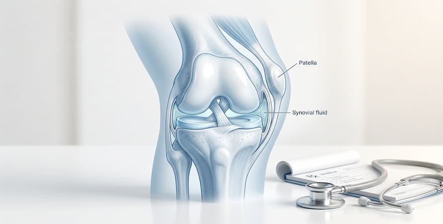

Everyone has a certain amount of synovial fluid in their joints. This fluid serves as a lubrication system that eliminates friction and helps to promote a somewhat well-oiled joint rotation.
Joint effusion[1] occurs when too much synovial fluid builds up around the joint region. When this happens in the knee, it’s referred to as water on the knee or swollen k
nee. This can be the result of infection, injury, or a medical condition.
Knee joint effusion can be extremely painful and lead to prolonged discomfort. So, if you suffer or experience joint effusion, it’s a good idea to be well informed so that you can make better and more educated decisions regarding your treatment plan.
In this article, we’re going to take a look at the knee joint effusion causes, different available treatment options, and how people can use preventative measures to minimize the chances of it occurring.
How Joint Effusion Develops

When you were younger, did you ever spend hot summer days filling up balloons with water? If so, do you remember how it felt when the balloons were tied? Superheavy with fluid, right?
This metaphor is similar to what occurs in our joints. When an injury transpires, it produces a fluid build-up that can begin to feel like a stiff water balloon. Knee joint effusion, more commonly referred to as water on the knee, happens when a surplus fluid begins to collect around the knee joint.
Joints are spaces where bones come together and meet, also known as articulations. When discussing knee joint effusion, the word “effusion” means an excessive fluid amount that collects within the articulation.
Normal joints contain synovial fluid, just in smaller amounts. Yet in certain cases, for example, arthritis, surplus fluid can collect, and the knee becomes swollen and puffy. Research[2] shows that 27% of people will experience joint effusion during their lifetimes, meaning that it’s not particularly rare.
The most common knee joint effusion causes are ligament injury, meniscus trauma, and arthritis.
Joints Most Commonly Affected by Effusion
While joint effusion can occur in any synovial joint in the body, four joints account for the vast majority of clinically significant effusion cases in adults. Each has its own characteristic presentation, most common causes, and diagnostic and treatment considerations.
Knee Joint Effusion
The knee is by far the most commonly affected joint, accounting for the majority of joint effusion presentations seen in clinical practice. This is not surprising given the knee’s combination of structural complexity, significant weight-bearing load, and vulnerability to both traumatic injury and degenerative disease. The knee joint contains the largest volume of synovial fluid of any joint in the body under normal conditions and can accumulate several hundred millilitres of excess fluid in significant effusion episodes.
Knee joint effusion is visually and physically detectable even in relatively mild cases. The characteristic puffy, ballottable swelling around the kneecap and in the hollows on either side of the patellar tendon is often the first sign a patient notices. The most common causes are osteoarthritis, ACL and meniscal injuries, rheumatoid arthritis, and gout. A specific complication of chronic knee effusion is the development of a Baker’s cyst, a fluid-filled bulge at the back of the knee that forms when excess synovial fluid cannot be reabsorbed and accumulates in the popliteal bursa.
Hip Joint Effusion
Ankle Joint Effusion
Shoulder Joint Effusion
Shoulder joint effusion occurs in the glenohumeral joint, the ball-and-socket joint between the humeral head and the glenoid fossa of the scapula. Like hip effusion, shoulder effusion is not visually apparent in mild cases and requires imaging for confirmation. It presents clinically as diffuse shoulder pain, restricted range of motion, particularly external rotation and abduction, and morning stiffness.
Common causes include rotator cuff tears (particularly large full-thickness tears), glenohumeral osteoarthritis, adhesive capsulitis (frozen shoulder), rheumatoid arthritis, and septic arthritis. Shoulder effusion in the context of acute trauma warrants assessment for haemarthrosis, blood within the joint, which can accompany significant rotator cuff tears or proximal humeral fractures and requires different management than inflammatory or degenerative effusion.
Causes of Joint Effusion by Category
Knee joint effusion[3] is commonly a sign of inflammation and can be classified as either noninfectious or infectious. Joint effusion produced by infection is known as septic arthritis, whereas an aseptic effusion is the result of injury or trauma.
1. Traumatic Causes

2. Inflammatory Causes
There are two kinds of arthritis:
Osteoarthritis – is also popularly referred to as “wear-and-tear” arthritis Autoimmune – such as rheumatoid arthritis, juvenile idiopathic arthritis, gout, and psoriatic arthritis, in which the immune system attacks joint regions
In cases of osteoarthritis, joint effusion predominantly affects the knee area and is most typically associated with joint damage. In autoimmune arthritis, joint effusion is most likely linked with acute flare-ups or chronic inflammation that originates as an immune response.
These immune responses are common in cases of autoimmunity and gout. The effusion is characterized and created by a collection of uric acid crystals in the joint space. Gout symptoms can appear quickly and severely, resulting in excessive synovial fluid in the joint area.
Beyond the osteoarthritis and autoimmune arthritis causes already described, several other inflammatory conditions can produce joint effusion. Reactive arthritis, previously called Reiter’s syndrome, is a sterile inflammatory arthritis that develops 1–4 weeks following a gastrointestinal or urogenital infection (most commonly Chlamydia trachomatis, Salmonella, Campylobacter, or Shigella). It characteristically affects the large joints of the lower limb, particularly the knee and ankle, producing effusion, pain, and swelling without active infection within the joint itself.
Psoriatic arthritis can affect any joint and frequently produces effusion, particularly in the fingers (dactylitis, diffuse sausage-like swelling of an entire digit) and the large joints of the lower limb.
3. Infectious Causes
One of the primary causes of knee joint effusion is infection, which can originate from a deep injury, wound, or a medical procedure. In the case of a blood-based infection known as systemic infection, it can take hold in the joint region, causing excess fluid and swelling.
When an effusion is caused by infection, knee joint effusion symptoms are intense and worsen quickly. Specific conditions increase the risk of septic arthritis; some of these include diabetes, joint replacement surgery, arthritis, older age, and a weakened immune system.
Septic arthritis represents the most dangerous cause of joint effusion and must be excluded whenever a monoarticular (single joint) effusion develops acutely, particularly with fever, elevated inflammatory markers, and no clear traumatic cause. The most common causative organism is Staphylococcus aureus, which accounts for approximately 40–50% of septic arthritis cases in adults. Neisseria gonorrhoeae is the most common cause in sexually active adults under 40 and should always be considered in this demographic presenting with an acute monoarticular effusion.
The clinical consequences of untreated septic arthritis are severe and rapid. Bacterial enzymes and the host inflammatory response can destroy articular cartilage within days, making same-day diagnosis and treatment initiation essential. Any joint effusion accompanied by fever, rigors, a very warm and red joint, or a markedly elevated white blood cell count requires emergency medical evaluation and aspiration for microbiological analysis before antibiotics are started.
4. Metabolic Causes
Two metabolic conditions deserve specific mention as causes of joint effusion because they are both common and frequently underdiagnosed:
Symptoms of Joint Effusion
The first symptom of knee joint effusion is the swelling of the knee region. This is tied to reduced and restricted mobility. The symptoms progress to cause pain and discomfort because the surplus synovial fluid within the knee produces significant pressure often felt behind the kneecap. A good way to judge the level of inflammation is by assessing the reddening, swelling, or heat coming from the knee.
Whatever the reason behind the fluid build-up, knee joint effusion symptoms tend to be similar, yet their severity will vary. Traditional symptoms of knee joint effusion[4] include:
- Stiffness – This limits a joint’s motion range or begins to restrict movement
- Swelling – This can range from mild discomfort to painful swelling
- Warmth and redness – These symptoms are commonly associated with inflammation
- Pain – The levels of pain can vary from a dull throbbing to an uncomfortable degree of pain that inhibits movement

Depending on the root cause of knee joint effusion, individuals may further experience the following symptoms as well:
- Malaise, chills, fever, and fatigue (infection is most likely present)
- Bleeding and bruising of the joint region (generated by an injury)
- Advanced muscle loss (this symptom stems from long-term arthritis, also referred to as atherogenic muscle inhibition)
A very commonly known complication of knee joint effusion is the appearance of a fluid-filled nodule within the joint space, more popularly referred to as a Baker’s cyst. This type of cyst occurs when the level of fluid build-up becomes excessive and cannot be absorbed or removed by the body naturally. Nevertheless, smaller Baker’s cysts do not cause symptoms or pain.
Distinguishing Inflammatory from Non-Inflammatory Effusion by Symptoms
A clinically important distinction that guides management is whether the effusion is inflammatory or non-inflammatory in character. Several symptom patterns help make this distinction before formal testing:
Non-inflammatory effusion (typical of OA and traumatic causes) is characterised by: mild-to-moderate swelling that develops gradually; pain that worsens with activity and improves with rest; morning stiffness lasting less than 30 minutes; minimal warmth or redness; and the absence of systemic symptoms such as fever or fatigue.
Inflammatory effusion (typical of RA, gout, reactive arthritis, psoriatic arthritis) is characterised by: more pronounced swelling and redness; morning stiffness lasting more than 30–60 minutes; warmth of the joint surface; systemic symptoms including fatigue and in some cases low-grade fever; and the pattern of affecting multiple joints rather than a single joint in isolation.
Septic arthritis produces the most severe and rapidly progressing symptoms: a single acutely swollen, intensely warm, red, and painful joint; high fever and rigors; markedly elevated inflammatory markers; and inability to bear weight on or move the affected joint. This is a medical emergency requiring immediate evaluation.
How Is Joint Effusion Diagnosed?
Joint Aspiration (Arthrocentesis)
Imaging Studies
Laboratory Analysis of Synovial Fluid
The synovial fluid white cell count (WBC) is the most diagnostically important laboratory result:
| WBC Count | Classification | Typical Causes |
|---|---|---|
| <200 cells/μL | Normal | – |
| 200–2,000 cells/μL | Non-inflammatory | OA, trauma, hypothyroid arthropathy |
| 2,000–50,000 cells/μL | Inflammatory | RA, gout, pseudogout, reactive arthritis |
| >50,000 cells/μL | Infectious (septic) | Bacterial septic arthritis |
| >100,000 cells/μL | Highly suspicious for infection | Emergency treatment required |
Crystal analysis under polarised light microscopy distinguishes gout (needle-shaped monosodium urate crystals, negatively birefringent) from pseudogout (rhomboid-shaped CPP crystals, positively birefringent), a critical distinction because both present similarly clinically but require different management.
Treatment of Joint Effusion

Drainage Or Aspiration
A medical professional uses a syringe to draw out the surplus synovial fluid, which can reduce pain.
Joint aspiration serves both diagnostic and therapeutic purposes. Therapeutically, removing excess fluid from the joint reduces the pressure within the joint space, providing immediate pain relief and improved range of motion. For large effusions, aspiration can remove 50–100 mL or more of fluid from the knee, producing dramatic immediate relief.
Aspiration is typically performed in combination with a corticosteroid injection for inflammatory causes. The steroid reduces the synovial membrane’s fluid production after the mechanical decompression. For gout and pseudogout, aspiration alone often produces significant relief as the crystal-laden fluid is removed, and the subsequent addition of a corticosteroid injection rapidly resolves the remaining inflammation.
Aspiration should be performed using sterile technique, either with anatomical landmark guidance or ultrasound guidance for deeper joints. Ultrasound guidance significantly improves accuracy, reduces the risk of incomplete aspiration, and is standard of care for hip and shoulder aspiration.
Medications
NSAIDs (Nonsteroidal Anti-Inflammatory Drugs): Oral NSAIDs, including ibuprofen, naproxen, and prescription-strength diclofenac and celecoxib, reduce the synovial membrane inflammation that drives excess fluid production. They are the most commonly used first-line pharmacological treatment for inflammatory and osteoarthritic joint effusion. Topical NSAIDs (diclofenac gel) applied directly to the affected joint provide a local anti-inflammatory effect with lower systemic absorption.
Colchicine: The first-line acute treatment for gout- and pseudogout-related joint effusion, most effective when initiated within the first 24–36 hours of an acute attack. It works by interfering with the inflammatory cascade triggered by crystal ingestion by neutrophils.
Urate-Lowering Therapies (allopurinol, febuxostat): For recurrent gouty effusion, long-term urate-lowering therapy reduces serum uric acid below the crystallisation threshold, preventing crystal formation and recurrent effusion episodes over time.
Antibiotics: The cornerstone of treatment for septic arthritis-related effusion. Empirical broad-spectrum antibiotics are initiated promptly after joint aspiration and adjusted based on culture and sensitivity results. Staphylococcal infections typically require 4–6 weeks of treatment; gonococcal infections typically respond to 7–14 days.
DMARDs and Biologics: For RA and other autoimmune arthropathies, disease-modifying treatment with methotrexate, hydroxychloroquine, sulfasalazine, or biologic agents (TNF inhibitors, IL-6 inhibitors) addresses the underlying immune dysregulation driving recurrent synovitis and effusion.
Injections
Surgery
Surgery for joint effusion is considered when conservative management, aspiration, and injection fail to control recurrent effusion, when significant structural damage requires repair, or when the underlying cause is inherently surgical.
- Arthroscopy may be performed to address specific structural causes of recurrent effusion, including meniscal tears, loose bodies within the joint, osteochondral defects, and pigmented villonodular synovitis (a benign but aggressive synovial proliferative condition that causes chronic bloody effusion).
- Synovectomy, surgical or arthroscopic removal of the thickened, inflamed synovial membrane, may be considered for chronic inflammatory effusion from RA or PVNS that has not responded to medical management.
- Joint replacement (arthroplasty), total or partial, may be considered for end-stage OA-related joint disease when symptoms remain severe despite appropriate conservative treatment.
Prevention and Long-Term Management of Joint Effusion
Frequently Asked Questions
Normally, knee injury recovery time lasts around 6 weeks. However, if surgery is required, the recovery time will increase and last anywhere between 8 weeks a year. The recovery time is dependent on many things, including the cause and severity of your effusion case.
An effusion is commonly the symptom of a condition or injury affecting the joint. In most cases, if the root cause is treated, the knee joint effusion will heal and go away. It's crucial to seek immediate medical attention if a joint effusion occurs for no reason or is paired with a high fever.
It's considered serious because it can be painful, especially when there is movement involved. And some cases of knee joint effusion, especially when paired with old-age or diabetes, can raise the risk of contracting septic arthritis.
Yes, the surplus synovial fluid build-up in the joint region can result in discomfort, stiffness, and pain.
Follow the advice outlined in this article and see your healthcare provider. Your doctor will assess your case and define a treatment protocol that will help get rid of your knee joint effusion.
Joint effusion and arthritis are related but distinct. Arthritis refers to joint inflammation or degeneration from a range of underlying conditions. Joint effusion is a symptom — the accumulation of excess synovial fluid — that commonly occurs as a consequence of arthritis. Not all arthritis causes significant effusion, and not all joint effusion is caused by arthritis. Traumatic injuries, infections, and metabolic conditions can all cause effusion in joints that have no underlying arthritic disease. In most cases of osteoarthritis and inflammatory arthritis, however, joint effusion is a recurring feature of the condition that worsens during active disease periods.
Joint effusion itself is not directly damaging in the short term, but persistent or recurrent large effusions can contribute to joint damage over time through two mechanisms. First, the elevated pressure within the joint from excess fluid compresses the articular cartilage and reduces nutrient delivery to this avascular tissue, potentially contributing to cartilage degradation. Second, the inflammatory mediators present in inflammatory effusion — particularly cytokines such as TNF-alpha and IL-1 — directly drive cartilage and bone erosion with repeated exposure. Septic arthritis-related effusion is the most destructive — bacterial enzymes can cause irreversible cartilage destruction within days without treatment.
Gentle exercise — particularly low-impact movement that maintains range of motion and periarticular muscle strength without loading the joint excessively — is generally appropriate for mild-to-moderate non-infectious joint effusion. Walking, swimming, and cycling are typically better tolerated than running or high-impact activity. High-impact exercise that provokes or worsens swelling should be avoided until the effusion has resolved. For acute or large effusions, initial rest followed by guided physiotherapy under professional supervision is the safest approach. Exercise with septic arthritis-related effusion is contraindicated — the joint should be rested until the infection is fully treated.
A Baker's cyst (popliteal cyst) is a complication of knee joint effusion rather than a separate condition. When the knee joint contains excess synovial fluid that cannot be reabsorbed, the fluid can be pushed through a natural weakness in the posterior joint capsule into the popliteal bursa at the back of the knee — forming the characteristic soft, fluid-filled bulge behind the knee that is a Baker's cyst. Treating the underlying joint effusion — by addressing its cause and aspirating the fluid — typically resolves the Baker's cyst as well. Isolated treatment of the Baker's cyst without addressing the effusion is rarely effective.
Seek emergency medical attention immediately for joint effusion accompanied by: high fever (above 38.5°C/101.3°F); a joint that is intensely hot, red, and severely painful; rapid onset of severe swelling within hours; the inability to bear weight or move the affected joint; or any systemic symptoms including rigors, confusion, or signs of sepsis. These features suggest septic arthritis — a medical emergency in which same-day joint aspiration, microbiological analysis, and antibiotic initiation are required to prevent irreversible joint destruction and life-threatening systemic sepsis.
Conclusion
Surplus fluid in the joint region is called an effusion, which affects more extensive joints such as the knee. A knee joint effusion occurs due to infection, injury, medical condition, or arthritis.
People find that after joint damage or serious injury, the knee will improve only to have fluid build-up again later. But, in most cases, the synovial fluid can be drained, and steps can be taken to address the root cause of the effusion.